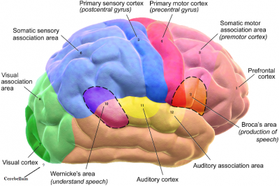
BRAIN
Safely inside the skull, the brain allows us to sense, think, learn, remember, and move. It also automatically regulates vital functions such as breathing. The cerebrum, the main part of the brain, has two halves, or hemispheres. The left hemisphere controls the right side of the body and is in charge of language, maths, and problem solving, while the right side controls the body’s left side and deals with creativity, music, and art. The cerebrum’s many tasks are carried out by its outer layer, or cortex. This has distinct areas that have different roles. Motor areas trigger movement; sensory areas deal with the senses, while association areas interpret information.
- PREFRONTAL CORTEX The most complex part of the cerebrum, the prefrontal cortex makes us what we are. It determines our personality and intellect, and enables us to reason, plan, create, and learn complex ideas, and have a conscience.
- BROCA’S AREA Named after Paul Broca, the 19th-century doctor who discovered it, Broca’s area is normally found in the left hemisphere. It plans what a person wants to say and sends instructions to muscles in the throat, tongue, and lips that produce speech.
- PREMOTOR CORTEX Learned movement skills, such as playing tennis, are controlled and coordinated by the premotor cortex. It tells specific muscles to contract either through the primary motor cortex or, in some cases, directly.
- PRIMARY MOTOR CORTEX Most movements we make are controlled by the primary motor cortex. Guided by information from the cerebellum and other brain parts, it sends instructions to muscles that move the skeleton instructing them when, and in what sequence, to contract.
- PRIMARY SENSORY CORTEX Receptors in the skin for touch, pressure, vibration, heat and cold, and pain send signals to the primary sensory cortex enabling us to feel those sensations. Our lips and fingertips have high concentrations of receptors, hence their sensitivity.
- SENSORY ASSOCIATION CORTEX Basic information about touch, pressure, and other skin sensations is passed on by the primary sensory cortex to the sensory association cortex. Here sensations are analyzed, stored, and compared with previous experiences. It enables us to identify objects by touch.
- PRIMARY VISUAL CORTEX When light hits the retina at the back of each eye; its light detectors send signals to the primary visual cortex. Here those signals are interpreted as basic shapes, colours, and movements before being passed on to the visual association cortex.
- VISUAL ASSOCIATION CORTEX This is where information from the primary visual cortex about seen objects is interpreted and compared with previous visual experiences. The visual association cortex identifies what we are looking at and where it is in space, enabling us to “see” it.
- CEREBELLUM The cerebellum is responsible for producing smooth, coordinated movements of the body. It analyzes incoming information about the body’s current position and movement, and then interacts with the primary motor cortex to precisely time muscle contractions.
- WERNICKE’S AREA Usually located in the left hemisphere, Wernicke’s area gives meaning to words that have been heard or read. Named after the German doctor Karl Wernicke, it has a direct link to Broca’s area enabling us to speak the words we hear or see.
- PRIMARY AUDITORY CORTEX When sounds are detected by the two ears they send signals to the primary auditory cortex. Here the loudness, pitch (whether high on low), and rhythm of sounds are identified. That information is passed on to the auditory association area.
- AUDITORY ASSOCIATION AREA Sounds are “heard” in the auditory association area. Using information received from the primary auditory cortex, it pieces together the complete sound, and, by comparing it with sounds stored in memory, identifies it as, for example, music, speech, or thunder.
Picture Credit : Google