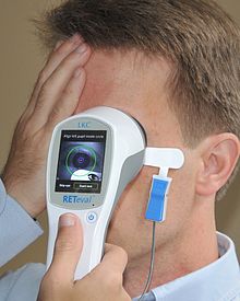
The recording of potential changes produced by the eye when the retina is exposed to a flash of light is called the electroretinogram, or in short ERG. Retina is a light-sensitive area at the back of the eye connected to the brain by the optic nerve. To record an ERG, one electrode is mounted on contact lens that fits over the cornea and the other is attached above the ear or forehead.
An ERG signal is more complicated than a nerve signal because it is the combination of the effects taking place within the eye. The general pattern of an ERG is shown in the figure. From the medical point of view, the ‘B’ wave is the most important because it arises in the retina. If a patient is suffering from ‘Retinitis pigmentosa’ the ‘B’ wave would be absent in his ERG because of the inflammation of the retina.
The recording of the potential changes due to eye movement is called electroculogram. In short, it is known as EOG. To record an EOG, a pair of electrodes is attached near the eye. An EOG can record horizontal and vertical movements of the eye. EOG also provides information about the orientation of the eye, its angular velocity, and its angular acceleration. Scientists have studied the effects of drugs on the eye movement. By an ERG, eye movements during sleep can also be studied. EOG is very rarely used in the routine check up for eye ailments.
Retina is an important part of the eye. It is a layer of special cells at the back of an eye ball and contains sensory cells capable of converting light into nervous messages that pass down the optic nerve to the brain. Images of the objects are formed on the retina. So with the help of an ERG, certain major diseases of the retina can be detected. Damage to the retina can cause blindness.


