Do you know what makes a chameleon change?


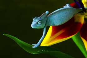
Chameleons are reptiles found predominantly in Madagascar, and some parts of Africa, Europe and Asia. There are over 150 species, in varying sizes and inhabiting rainforests, deserts, etc. While some reside close to the ground, many of them live in trees. And that would explain their grouped toes for climbing and prehensile tails curled around branches for balance. But what sets chameleons apart from most creatures is their colour-changing ability.


Myriad colours, but…


Yes, chameleons do change colour and this may help in camouflage. But most of them already have skin colour that help them blend well with their surroundings. For instance, it is said that tree-dwelling chameleons are in shades of green while their cousins in deserts are more in shades of brown. So it’s a misconception that they can match the colours of their background. Reports say these reptiles predominately change colour for defence, to communicate. Attract mates, display anger, fear, threat, etc., and as a response to changes such as light or humidity in their surroundings.


Here’s how it’s done. The chameleon skin has a few layers. The outermost is said to be transparent. The layer beneath this has special iridescent cells with different colour pigments that also reflect light. The reptile has the ability to change the arrangement of these cells – “by relaxing or exciting the skin”.


All-seeing eyes and long tongues


In addition to a colour-changing skin, they have another rare ability – eyes that move independently of each other, giving these reptiles the chance to look in two different directions simultaneously. When they want to look at something specific, both eyes focus on that object together. Not just that, their cone-shaped eyes rotate, giving them a 360-degree view of their surroundings. Another interesting aspect of the chameleon is its tongue. The sticky tongue is said to be almost twice the length of its body. When it spots its prey, the tongue shoots out with incredible speed, acts as a suction cup and pulls the prey in before the unsuspecting victim has any time to react!


Picture Credit : Google


What are the blood types?


There are four main types of blood group – A, B, AB, and O. The group mostly depends on special makers on the surface of red blood cells, called antigens. These help the body to identify blood cells that do not belong to you. Patients who need a blood transfusion must receive the right blood group, or the body will reject the donated blood, making them even more unwell.


Type A


This can be donated to those with type A or AB blood groups. The surface of the red blood cells contains A antigen, and the plasma has anti-B antibody. Anti-B antibody would attack blood cells that contain B antigen.


Type B


This can be donated to those with type B or AB blood. The surface of the red blood cells contains B antigen, and the plasma has anti-A antibody. Anti-A antibody would attack blood cells that contain A antigen.


Type AB


This blood can only be donated to other people with the AB blood type. The red blood cells have both A and B antigens, but the plasma does not contain anti-A or anti-B antibodies. Individuals with type AB can receive any ABO blood type.


Type O


These blood cells have no antigens so can be donated to people of any blood group. The plasma contains both anti-A and anti-B antibodies, but the surface of the red blood cells does not contain any A or B antigens. Since these antigens are not present, a person with any ABO blood type can receive this type of blood.


Picture Credit : Google


What is the life cycle of red blood cells?


All blood cells are made in red bone marrow. Almost all the bones of young people contain red bone marrow. For adults, it is only found in the skull, ribs, shoulder blades, hips, and the ends of long bones.


Cell cycle


A red blood cell lives for up to 120 days before being swallowed by a type of white blood cell, called a macrophage, in the liver or spleen.


New blood cells are made by red bone marrow. A new red blood cell is released into the blood stream. The worn-out red cell is digested by a macrophage. Waste from the blood cell is removed. Useful parts of the old blood cell are recycled.


Picture Credit : Google


How is blood transported in the body?


Blood is constantly moving oxygen, nutrients, proteins, and waste products around the body. Some of these help cells grow and function, others are converted into new substances, and the rest are removed from the body.


Oxygen carrier


Red blood cells contain a protein called haemoglobin. Oxygen that enters the blood in the lungs attaches to this haemoglobin and is later released into the body’s tissues. It is haemoglobin that gives blood its red colour – and the more oxygen haemoglobin carries, the brighter red it becomes.


When red blood cells pass through the lungs, haemoglobin binds with oxygen. Red blood cells carrying oxygen travel where they are needed in the body. When the red blood cell arrives at its destination, the haemoglobin releases oxygen.


Transport superhighway


The bloodstream provides an efficient delivery service, delivering essential fuel and oxygen to cells, and at the same time taking away waste and toxins to keep cells and tissues healthy.


Oxygen


Red cells pick up oxygen in the lungs and deliver it to all the cells in the body.


Nutrients


Nutrients enter the blood from the digestive system and are delivered around the whole body.


Water products


Water is delivered to the liver for recycling or to the kidneys to be made into urine.


Hormones


The blood delivers chemical messengers, called hormones, to specific destinations.


Body defences


White cells are carried to fight germs. Platelets are delivered to wound sites.


Carbon dioxide


Carbon dioxide made by cells is delivered to the lungs to be breathed out of the body.


Picture Credit : Google


What is in blood?


Blood consists mostly of fluid called plasma, together with three types of blood cell – red blood cells, white blood cells, and platelets. They all have different jobs to carry out inside the body.


Blood breakdown


Plasma is made of water with substances dissolved in it, including salts, nutrients, and hormones. Red blood cells take oxygen to cells and remove carbon dioxide. White blood cells hunt and kill bacteria and viruses, while platelets repair damage by plugging a wound and helping blood to clot (thicken).


Plasma


When a blood vessel tears, platelets and plasma proteins work together to stop blood loss. Pale yellow plasma is 91% water.


Platelets


Rounded platelets become spiky when blood clots. Platelets, also called thrombocytes, clump and form a plug in the damaged area. The proteins form threads called fibrins to complete the platelet plug, or clot.


White blood cells


These cells are the largest in the blood. White blood cells, also called leukocytes, are the disease-fighting components of blood. They account for just 1% of circulating blood but multiply during infection or inflammation. There are five types of white blood cells: neutrophils, eosinophils, basophils, lymphocytes, and monocytes. Neutrophils are the most abundant, comprising 60% to 70% of all white blood cells.


Red blood cells


Just under half of blood is made up of red blood cells. Red blood cells, also called erythrocytes, make up most of that 45%. Their primary function is to transport oxygen from the lungs to the cells of the body. Red blood cells are disc-shaped. They are flexible and bioconcave—flat and round with depressed centers.


Picture Credit : Google


What is the function of blood?


Blood circulates endlessly through the human body to keep it alive. This fluid contains trillions of cells and countless chemicals, all floating in watery plasma. Blood is pumped by the heart through a network of blood vessels to deliver nutrients, oxygen, and other essential substances to cells. Blood also transports waste; helps keep the body temperature steady, and fights germs.


Blood flows into the kidneys through the renal arteries and out through the renal veins. The kidneys filter substances such as urea, uric acid, and creatinine out of the blood plasma and into the ureters. The liver also removes toxins from blood. During digestion, it cleans blood that has been enriched with vitamins before sending it back out to the rest of the body.


Blood vessels expand and contract when they react to outside organisms, such as bacteria, and to internal hormone and chemical changes. These actions move blood and heat closer to or farther from the skin surface, where heat is lost.


Picture Credit : Google


What is the structure of blood vessels?


The walls of both arteries and veins are made up of three main layers – a tough outer coating, a wall of muscle, and a smooth, inner lining. Arteries have a thicker middle layer of muscle to control the flow, or pressure, of blood. Pressure needs to be high enough to push blood around the system, but not so strong that it damages delicate capillaries.


Wall of muscle


Arteries’ muscular walls stretch to cope with the high-pressure pulses of blood pumped out by the heart. The muscle contracts to make the artery narrower and reduce blood flow, and relaxes to widen it and allow blood to flow more freely.


Artery:


Blood


Blood is made up of three types of cells floating in yellowish liquid called plasma.  Arterial blood has just passed through the lungs and is ready to boost oxygen to sustain the peripheral organs. 


Inner layer


The smooth inner lining lets blood flow easily. The innermost layer, the tunica intima (also called tunica interna), is simple squamous epithelium surrounded by a connective tissue basement membrane with elastic fibers. 


Membrane


A thin, protective covering surrounds the inner layer. The vascular basement membrane is a dynamic, self-assembled layer of proteins, glycoproteins, and proteoglycans formed by and enveloping endothelial cells and pericytes of blood vessels. 


Muscle layer


Muscular arteries contain more smooth muscle cells in the tunica media layer than the elastic arteries. 


Elastic layer


Elastic arteries are those nearest the heart (aorta and pulmonary arteries) that contain much more elastic tissue in the tunica media than muscular arteries. These layers allow the artery to stretch and bounce back into shape.


Capillaries


Capillaries, the smallest and most numerous of the blood vessels, form the connection between the vessels that carry blood away from the heart (arteries) and the vessels that return blood to the heart (veins). The walls of capillaries are only one layer of cells thick, enabling gases to pass easily through them.


Outer layer


The artery wall is made of tough but flexible collagen. This layer is connective tissue with varying amounts of elastic and collagenous fibers.


Vein:


Valve


Valves in the veins make sure the blood flows one way only. Valves also help blood travel back to the heart against the force of gravity.


Muscle layer


The layer of muscle is thinner than an artery’s.


Picture Credit : Google


What is the function of capillary and vein valve?


Capillary connection


Capillaries connect arteries and veins. The wall of each capillary is formed from an ultra-thin layer of flattened cells. This lets gases and nutrients pass easily through the wall. Some capillaries also have pores, called fenestrations, to make the exchange even quicker.


Vein valve


The long veins in the leg have valves to make sure that blood travels up towards the heart and doesn’t fall back down to the feet. When the muscles around the vein contract, they open the valve and push blood upwards. When the surrounding muscles relax, the valves close to stop the blood flowing back down.


Picture Credit : Google


What are blood vessels?


Pumped by the heart, blood circulates around the human body through millions of blood vessels. These deliver oxygen and other essential substances to the body’s cells and tissues.


The three types of blood vessels are arteries, veins and capillaries. Arteries carry oxygen-rich blood away from the heart. Veins carry oxygen-poor blood back to the heart. These two networks are linked by the smallest blood vessels, capillaries. Oxygen seeps through their thin walls into cells and tissues, while carbon dioxide goes in the other direction, from cells to capillaries.


The inner surface of every blood vessel is lined by a thin layer of cells known as the endothelium. The endothelium is separated from the tough external layers of the vessel by the basal lamina, an extracellular matrix produced by surrounding epithelial cells. The endothelium plays a critical role in controlling the passage of substances, including nutrients and waste products, to and from the blood. Under certain circumstances, tissues may grow new blood vessels, a process known as angiogenesis. Angiogenesis plays an important role in the replacement of damaged tissue but also occurs under abnormal conditions, such as in tumour growth and progression.


Picture Credit : Google


How the heart works?


Although a heartbeat lasts just one second, it has three stages. The rate at which the heart beats is controlled by a pacemaker located in the wall of the right atrium, which sends electrical signals to all parts of the heart.


Blood flows into the atria


The heart muscle is relaxed and blood enters the upper left and right heart chambers (atria).


From atria to ventricles


The two atria contract and squeeze blood into the chambers below them (ventricles).


Blood leaves the heart


Lastly, the ventricles contract and force blood either to the lungs or around the body.


Picture Credit : Google


What is inside the heart?


The heart is really two pumps in one, working in a continuous cycle. The right side pumps blood to the lungs, while the left side receives blood back from the lungs, then sends it round the rest of the body.


The heart pumps out about a cupful of blood about 70 times a minute, speeding up when necessary to meet body cells’ increased demand. Over a lifetime, the heart beats more than 2.5 billion times without resting.


Each side of the heart has a small upper space, called an atrium, and a larger space below called a ventricle. During each heartbeat, blood is pumped from atrium to ventricle, then out of the heart. Valves open and close to make sure that the blood only flows in one direction.


Aorta


The aorta is the first segment of the systemic arterial circulation, originating directly from the left ventricle of the heart. The aorta is the body’s largest artery.


Pulmonary artery


The pulmonary arteries and the pulmonary veins are the vessels of the pulmonary circulation; which means they are responsible for carrying the oxygenated blood to the heart from the lungs and carrying the deoxygenated blood from the heart to the lungs. This major blood vessel delivers oxygen-poor blood to the lungs.


Pulmonary veins


Oxygen-filled blood is carried from the lungs to the heart by these veins. here are 4 total pulmonary veins—with 2 pulmonary veins coming from each lung, left and right—that empty into the left atrium of the heart


Pulmonary valve


The pulmonic valve is one of two valves that allow blood to leave the heart via the arteries. This prevents blood flowing back into the right ventricle from the pulmonary artery.


Tricuspid valve


The tricuspid valve, or right atrioventricular valve, is on the right dorsal side of the mammalian heart, at the superior portion of the right ventricle. This controls blood flow on the right side of the heart.


Right atrium


The right atrium (RA) is one of the four chambers of the human heart, and is the first chamber to receive deoxygenated blood returning from the body. Oxygen-poor blood from the body flows into this chamber.


Left atrium


The left atrium is one of the four chambers of the heart, located on the left posterior side. Its primary roles are to act as a holding chamber for blood returning from the lungs and to act as a pump to transport blood to other areas of the heart. Oxygen-rich blood from the lungs flows into this space.


Mitral valve


This controls blood flow on the left side of the heart. The mitral valve and the tricuspid valve are known collectively as the atrioventricular valves because they lie between the atria and the ventricles of the heart.


Septum


The right and left sides of the heart are divided by this wall of muscle. Histological septa are seen throughout most tissues of the body, particularly where they are needed to stiffen soft cellular tissue, and they also provide planes of ingress for small blood vessels. 


Left ventricle


The left ventricle is one of four chambers of the heart. It is located in the bottom left portion of the heart below the left atrium, separated by the mitral valve. Oxygen-rich blood travels from this chamber to the aorta.


Right ventricle


This chamber pumps oxygen-poor blood to the lungs. The right ventricle is one of the heart’s four chambers. It is located in the lower right portion of the heart below the right atrium and opposite the left ventricle.


Pericardium


The pericardium is a double-walled layer of tissue surrounding the heart. This tough, double-layered “bag” around the heart keeps infections out and stops the heart from expanding too much when blood flows into it.


Heartstrings


The heartstrings of the tricuspid valve sit between the right atrium and right ventricle. These tough cords stop the valves from turning inside out when the ventricles contract.


Picture Credit : Google


What is the blood supply of heart?


To keep the heart beating, the cells need a constant supply of fuel and oxygen. This is delivered by the coronary blood supply – the heart’s own network of blood vessels, which penetrate the heart’s walls to reach the muscle.


Healthy heart


Than angiogram is an intricate network of blood vessels branching off the main arteries.


Tight-squeeze


The heart sits in the chest, surrounded by the ribcage and between the two lungs, which take up most of the rest of the space in the chest cavity.


Heart rate


The heart rate is the number of beats per minute (bpm) that the heart makes. A person’s average heart rate varies according to different factors, such as age, gender, and level of fitness.


Picture Credit : Google


What is the anatomy of heart?


A healthy adult’s heart is about the size of a clenched fist. It sits in the thorax (chest) and in most people the tip points towards the left side of the body.


Aorta


The aorta is the first segment of the systemic arterial circulation, originating directly from the left ventricle of the heart. The body’s main artery carries blood from the heart to the rest of the body.


Superior vena cava


The superior vena cava (SVC, also known as the cava or cva) is a short, but large diameter vein located in the anterior right superior mediastinum. This large vein returns oxygen-poor blood from the upper body to the heart.


Pulmonary artery


The pulmonary arteries and the pulmonary veins are the vessels of the pulmonary circulation; which means they are responsible for carrying the oxygenated blood to the heart from the lungs and carrying the deoxygenated blood from the heart to the lungs. Blood is carried to the lungs by this vessel.


Coronary artery


The heart’s own blood supply is delivered by this artery. The right coronary artery supplies blood mainly to the right side of the heart. The right side of the heart is smaller because it pumps blood only to the lungs.


The left coronary artery, which branches into the left anterior descending artery and the circumflex artery, supplies blood to the left side of the heart. 


Muscle structure


 Heart muscle shows that the structure is a network of interlocking fibres. The oval discs are mitochondria, which supply the muscle cells with the energy they need.


Pericardium


This tough, double-layered “bag” around the heart keeps infections out and stops the heart from expanding too much when blood flows into it. It also allows the heart to beat without rubbing against other organs.


Picture Credit : Google


What is the function of heart?


The heart is the engine at the centre of the body’s circulation system. It starts working even before we are born – and from the on, it beats constantly, throughout our lives.


Made from a special kind of muscle not found anywhere else in the body, the hardworking heart contracts and relaxes about 70 times every minute. This rhythmic pumping pushes essential blood out to the body, then fills the heart up again, ready for the next beat.


The performance of the heart could now be easily monitored when any cardiovascular problem or disorder is suspected. For instance, a regularly abnormal heartbeat or beats per minute are characteristic of a heart-related illness. This is because a heartbeat is a manifestation of the oxygen-reloading process in the heart that is made up of two phases.


Picture Credit : Google