How surgeons smooth away the wrinkles?


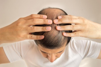
As the skin ages, some of the subcutaneous or underlying fat which supports and pads it dissolves away. And one of the skin-s main constituents, called collagen, loses its ability to retain moisture, making the skin less elastic and drier. The result is sagging skin and wrinkles.


Most people accept wrinkles as part of growing older. For others, particularly those in the public eye like entertainers and politicians, ageing skin can be a problem. The only answer is cosmetic surgery.


There is more to cosmetic surgery than a face-lift — which, as its names suggests, means pulling the skin up over the face. Its cosmetic effects are, for the most part, restricted mostly to the chin and neck. Wrinkles around the eyes the side of the, nose, and across the forehead have to be dealt with in separate operations, such as an eyebrow or forehead lift, or a nasal fold removal. In blepharoplasty, excess loose skin is removed from the upper and lower eyelids.


Minor nips and tucks arc clone under local anaesthetic, bat a face-lift is a major


operation, and is usually done under general anaesthetic. The surgeon first makes an incision into the skin around each ear. He starts the cut well within the hairline above the ear, and continues it around the bottom of the ear and then up behind it. The cut is then taken horizontally towards the back of the head. Most of the cut is within the area covered by hair, so that the scars will be hidden.


Once the cuts are made, the surgeon carefully separates the skin below the line of the cut from the underlying fatty layer. He then pulls the loose skin towards the back of the head. The thin layer of muscle tissue in the neck is lifted and tightened. The excess skin is cut off and the incision sewn up.


 It often takes two to three weeks to recover from the slight inflammation of the face caused by the operation. The scars, which can be camouflaged by make-up a week after the operation, fade in time.


No face-lift retards ageing permanently. The ageing process continues from the time of the operation at the normal rate. More face-lifts can be performed on the same person but there is always a limit, because each time the surgeon removes more skin. When the skin is stretched to its tightest limit without hindering normal functions, such as smiling, there is no excess available and further operations become impossible. Not all operations are a success and some people have been left with badly scarred faces.


Picture Credit : Google


What happens in a heart transplant?


When a heart becomes available, a suitable recipient is quickly located and told to get to the hospital immediately. At the same time, a combination of police, ambulance and helicopters race the donated organ to the hospital. A heart may travel hundreds of kilometres from donor to recipient, sometimes across international boundaries. But to save time, the European computerized system, Euro transplant, tries to locate recipients who live as close to the donor as possible.


To prepare a patient for a heart transplant, the surgeon cuts into the chest and ties off the blood vessels leading to and from the recipient's heart. The recipient's blood supply is then redirected through the heart-lung machine, which replaces the function of the patient's own heart and lungs. The faulty heart is taken out, and the new organ is placed in the space. The new heart is then connected to the major veins and arteries before the recipient's blood is diverted through the new organ. The surgeon then sews up the chest and the operation is complete.


Picture Credit : Google


How a pacemaker helps a heart patient to lead a normal life?


The human heart beats 3000 million times in an average lifetime, pumping the equivalent of 48 million gallons (218 million litres) of blood around the body.


The regular rhythm — on average, 72 beats each minute — is controlled by the sino-atrial node, a tiny rounded organ located in the top left corner inside the heart. This is the heart's natural pacemaker, which sends electrical impulses to the tissues. The heart contracts and heart's expands in response to these impulses, producing the heartbeat.


Occasionally, the heart's electrical con-ducting system can be disturbed by illness, such as angina or a heart attack. Sometimes it just fails completely. If this happens, the heart can be stimulated electrically to continue beating regularly.


If the heart stops it can sometimes be restarted with an electrical shock from a machine called a defibrillator. If the normal beat does not resume immediately, some-times a temporary pacemaker can be fitted outside the body — it is usually strapped to the waist. For those suffering from other irregularities of the heart beat a pacemaker is surgically placed inside the body, implanted in the chest.


 All pacemakers, inside and outside the body, work in the same way. An electrode on wire, called the end of a pacing lead, is attached to the wall of the heart's right ventricle (chamber), either directly through the chest, or threaded through a vein. The electrode is powered by the pacing box, a miniature generator operated by lithium batteries. Modern pacemaker batteries last at least five years, and some last up to 12 years.


 Powered by the pacing box, the electrode produces electrical impulses which stimulate the sino-atrial node and make the heart beat. The pacing box is set to maintain the intervals of the impulses at a given rate, usually one beat per second, which is a little slower than the average heart rate. However, the box functions only when the heart is not producing its own electrical impulses at the correct intervals. It is sensitive enough to detect these delays and by filling in the gaps, maintains a normal rhythm. Some models include a radio transmitter and receiver, which means that a doctor can adjust the rate of the pacemaker from outside the patient's body.


The first successful pacemakers were used by Dr Walter Lillehei, a cardiac specialist at the University of Minnesota, USA, in the late 1950s. They consisted of an electrode on a wire fed to the heart through the chest and attached to a battery pack strapped around the waist. The pack was about the size of a cigarette packet. Although the system was convenient because no surgery was needed to replace the batteries, the opening in the chest for the wire repeatedly became infected. External pacemakers are now used for temporary heart problems only, or until an internal pacemaker can be fitted.


The pacing box of the most commonly used internal pacemaker is about the size of a matchbox and weighs no more than 25g. It is usually made of lightweight titanium.


The box is implanted in the body, usually just inside the skin of the chest wall. It must be in the best position for threading the tube through the large vein to the heart and attaching the electrode, which is the size of a match head, to the heart wall. The body does not reject it because it is not living material.


The implanting operation is done while the patient is under general anaesthetic, but surgery to replace the batteries can usually be done with only a local anaesthetic.


 A person wearing a pacemaker needs to be examined by a doctor frequently to make sure that it is functioning properly. Also, some wearers have to take care that their pacemakers are not affected by certain electrical circuits, such as magnetic detectors in airports or libraries.


New electronic technology may produce even smaller pacemakers which can be attached to the heart wall, eliminating, wires and large battery packs, although!they are still powered by batteries.


 Another development is the rate-responsive pacemaker, which is sensitive to the patient's activity. Instead of providing at impulse once a second, it will increase the impulses when he is active and slow them down when he is resting — like the heart, natural pacemaker.


Since the First successful pacemaker developed, more than 5 million people with serious heart disease have been helped to live more comfortable and active lives.


Picture Credit : Google


How do anaesthetics numb pain?


Less than 150 years ago, surgery was performed without any anaesthetic. A patient was held down by strong men as he battled to escape from the pain of the surgeon's knife. Surgeons even resorted to stupefying their patients with alcohol, knocking them unconscious, or freezing the part to be operated on with ice.


The first time an anaesthetic was used was on March 30, 1842, in Jefferson, Georgia, USA, when Dr Crawford Long removed a tumour from the neck of James Venable, who first inhaled ether. But it was only following William Morton's public demonstration in Boston of the extraction of a tooth under ether, in 1846, that ether became widely adopted as an anaesthetic.


At around the same time in the United States, nitrous oxide, also known as laughing gas and used as a music hail entertainment, was being inhaled as an anaesthetic for dental surgery. In Britain. Research was being done on the uses of chloroform, particularly to relieve the pain of childbirth. Without these early attempts at the use of anaesthetics, many of today's surgical procedures would not be possible. Now, major operations, such as heart trans; plants, cosmetic surgery and removal,01 cancer, are possible without pain. But just, how do anaesthetics allow people to slip off into a world where pain does not exist?


Anaesthesia derives from the Greek word for 'lack of feeling'. All anaesthetics induce this condition by blocking the of pain signals to the brain. However, how they actually work is not yet fully understood.


Anaesthetics take two forms — general, which put the patient 'to sleep', and local, which affect only part of the body.


Loss of sensation, ox or analgesia may be provided by nitrous oxide, not put the patient to sleep. It may cause mental or physical excitement. Sleep is usually induced by an injected barbiturate. The muscles are then relaxed with a neuroblocker, or muscle relaxant, such as curare.


During surgery, the patient is watched so that any changes in circulation; so that any changes in circulation, breathing or kidney function which may result from the anaesthetic can be regulated.


Local anaesthetics are given as an injection to remove all sensation from and a localised area. The patient is conscious can cooperate with the surgeon.


There are three principal uses of local anaesthetic. Topical anaesthetics remove the sensation from nerve endings in mucous membranes such as those in the eye, the nose and the mouth. They are used, for example, to remove a foreign object from the eye. Nerve-block anaesthetics are injected into a nerve to anaesthetise a small area, for example, to enable a tooth to be extracted. Other anaesthetics are injected into a large nerve group to numb a larger part of the body, such as an arm.


Atoms that transmit pain


A clue to the way general anaesthetics work comes from research into local anaesthetics. These are known to interfere with the way nerve impulses are transmitted a along the nerve fibres. Sodium and potassium atoms play an important pa sending these impulses to the brain. If you stub your toe, for example, the sodium and potassium atoms pass in opposite directions across the membrane of the nerve cell causing the next cell to do the same and so on until the signal reaches the brain, when you feel pain. But local anaesthetics stop the atoms from passing in and out of the nerve cell, so no pain signal reaches the spinal cord.


 Scientists think that general anaesthetics may cause unconsciousness by suppressing the activity of certain enzyme in the nerve cells, or changing the prop of the nerve-cell membranes, or even by interacting with water molecules in the brain to form small crystals which affect the path of a signal along a nerve cell. Research. Continues into the exact -mechanism, but what is certain is that without anaesthetics a great deal of surgery could never he performed.


Picture Credit : Google


How surgeons do bloodless operations with just a beam of light?


Until laser surgery was invented in 1963, someone with a growth, a cancer or a cataract, needed a major operation to have it removed. Now, laser beams can be used in 'bloodless' operations to remove growths and even repair tissues, without cutting, less painfully and more safely.


 In laser surgery to remove a growth from the throat, for example, a small tube, or endoscope, is passed down the patient's throat, under local anaesthetic. A laser beam is directed down the tube along an optical fibre and is focused on the growth. All laser surgery works on this principle of passing light down an optical fibre. The beam is a form of light which carries a great deal of energy. The energy is absorbed by the tissues of the growth, or the skin tissues to be removed, which become hot. By controlling the heat intensity, doctors can burn off — literally vaporise — unwanted cells.


In this way, laser beams are used to cut away cancers, vaporise the dyes in tattoos or get rid of birthmarks.


Another use of laser beams is to heat tissues sufficiently to `weld' them together — to stop blood vessels bleeding, for example. The operation might be per-formed on a patient who is bleeding from a stomach ulcer.


 The wavelength of the laser beam affects the way in which tissues respond to it. Lasers that use carbon dioxide produce beams of light which are absorbed by tissues at a depth of only 0.1mm. This means that they can be used to make fine cuts in tissue, as a sort of 'laser scalpel'. Such precision cutting might be used when making incisions in the cornea of the eye to correct defects in sight, or in removing throat tumours.


 Lasers using a metal-based chemical called neodymium produce light which is absorbed by a greater depth of tissue, making it useful to destroy cancers.


Those lasers that use the gas argon produce a distinctive blue-green light, which is absorbed by haemoglobin — the chemical in the blood that gives it its red colouring. Argon beams can therefore be used where haemoglobin levels are high,, in birthmarks.


A further benefit of laser beams is that they allow doctors to reach areas of the body previously hard to get at with a scalpel and to perform operations that were impossible before: to rid arteries of block. Ages of fatty deposits; to sew back detached retinas; to cut a hole through a cataract in a lens and so restore vision; and to cure cancer of the cervix.


Picture Credit : Google


How do doctors perform microsurgery?


On Christmas Eve in 1980, Beatrice Ramos threw herself and her 13-month-old son, Vladimir, under a subway train in New York. Both were badly hurt. Vladimir's right foot and left leg were injured beyond repair. But to spare him from having two false limbs, surgeons at Bellevue Hospital performed a pioneering operation in which they attached his left foot to his right leg.


Only ten years earlier, such an operation would have been thought impossible. Now, operations to save limbs are much more common.


Microsurgery involves working on the tiniest structures in the human body, such as nerve fibres, veins and fine arteries. When sewing back a severed part of the body, it is not sufficient simply to sew it on. Without connecting blood vessels the part would die from lack of oxygen, and if the nerves were not connected, it would have no nervous stimulation and would be useless.


Since the structures involved are so fine — an artery in a finger is about I/16in (1-2mm) wide and a nerve fibre varies from .002mm to .02mm — microsurgery is possible only with high-powered microscopes. These instruments have a magnification from x 6 to x 40, allowing surgeons to see the tiny structures that need joining up. Micro-scopes with two or three heads have been developed, which allow more than one surgeon to work at the same time.


 When stitching nerves, surgeons have to make sure that they join matching bundles. They are usually identified before surgery.


The surgeon works with a needle which is only 50 microns (.05mm) thick, with 18 micron (nearly .02mm) nylon thread.


When stitching two blood vessels together. A surgeon normally uses a method known as triangulation. Three stitches are made 120 degrees apart at the end of the blood vessels, and then the surgeon sews all the way around their circumference, a third at a time.


It can take 15 to 30 minutes to stitch one vein to another. Stitching back a hand can take 19 hours.


Sometimes blood vessels can be joined together without intricate sewing. By using electrical probes to heat up the severed ends the surgeons can literally weld them together.


After surgery, physiotherapy is essential to restore the replanted limb to working order. For a replanted hand it takes about 200 days for the nerve and blood vessel tissues to regenerate. It takes longer. However, for the part to function normally.


 Apart from repairing injuries, micro-surgical techniques can be used for a host of other problems. Eye operations, for example, involve microsurgery. An eye operation called 'radial keratotomy', which was pioneered by Russian surgeons, can sometimes cure short sight. The surgeon makes a number of slits radiating from the centre of the cornea, the surface of the eye. The cuts change the shape of the cornea, which alters the distance between the front of the eye and the retina, bringing objects into focus which previously was not.


Brain surgeons use operating micro-scopes to place their instruments with much greater precision, so improving the chances of success in removing tumors. The microscopes enable surgeons to re-move the tumor without cutting away any normal brain tissue.


Picture Credit : Google


Why carbohydrates are important for athletes?


There is one dietary regime which has been shown to boost athletes' energy levels significantly. Known as carbohydrate loading, it increases the level of glycogen in the muscles. Glycogen is a form of glucose which is broken down to release energy. By building up the amount of glycogen they contain, the muscles can work hard for longer.


 The programme usually starts a week before a competition. On the seventh day before the event, preferably in the evening, the athlete performs a strenuous training routine to deplete the glycogen stored in his muscles, and eats a low-carbohydrate meal. During the following three days he trains less and continues with low-carbohydrate meals.


On days three and two the athlete eats a high-carbohydrate diet and eases the training further.


On day one the carbohydrate intake is increased again, and the athlete rests, in preparation for the event the following day.


The principle behind this programme is that when a high level of carbohydrates is introduced to muscles low on glycogen, the muscles overcompensate and take in a higher than normal level of glycogen over a short period. It is these surplus stores that the athlete draws on during his event, which keep him going longer.


 Following this dietary routine, some top-ranking marathon runners have found that their performances have improved significantly.


Picture Credit : Google


How people survive a bolt of lightning?


An American park ranger, Roy Sullivan, who lived in Virginia, was reputedly struck by lightning more times than anyone else in the world. He was first hit in 1942, and received his seventh strike on June 25, 1977. On each occasion Sullivan was injured. He first lost as toenail, then had his eyebrows burnt off, his shoulder was seared, his hair was set alight and his legs were burnt. But he survived.


Lightning can travel between 100 and 1000 miles per second (160 to 1600km per second), and the temperature it generates can reach 54,000ºF (30,000ºC), six times hotter than the surface of the sun. So hat is remarkable is that many people hit by this awesome force do not die, although they are usually burnt quite badly.


Lightning is a spark of electricity caused by the difference in energy between the clouds and the surrounding air or the ground. in cloud-top ground lightning the energy seeks the shorted route to earth, which may be a person out in the open.


This shortest route could be through the shoulder, down one side of the body, through the leg and into the ground. on the way it will cause pain, shock and burns, but the bulk of its energy is discharged into the ground. as long as the lightning does not pass across the heart or spinal column, the victim will probably not die. But if the forces crosses the heart, there is a great risk that it might be damaged , or stop, resulting in instant death.


Picture Credit : Google


How doctors can sometimes cure baldness?


More than 90 per cent of men succumb to some degree of baldness. And some women, particularly after the menopause, also find that their hair thins and recedes. The problem is entirely genetic – bald fathers have sons who are likely to become bald. And it doesn’t just affect humans either: monkeys become bald as well.


The exact genetic code that causes baldness and thinning still eludes researchers, but they know it has something to do with male sex hormones called androgens. These hormones suppress the activity of certain hair follicles on the scalp, so that the life span of their hair that grows from them is reduced. Normally, a hair allowed to grow uncut will last from two or six years. But as baldness sets in, the hair in some areas of the head falls out more often. The overall effect is that the hair in those places gets thinner and shorter, until it is reduced to fuzz.


Men have more androgen than women, which is why more men that women suffer from baldness.


One solution is baldness is a ‘scalp transplant’, which is carried out by a cosmetic surgeon. But only some people are suitable. Their baldness should be stable, that is, not getting worse each year, and should occur mainly at the front and top of the scalp. The remaining hair needs to be dark to hide the effects of the surgery. It also needs to be healthy and abundant because it is this hair that will be transplanted. In hair transplant surgery there are no donors, the patient’s existing hair is simply redistributed.


First the patient’s hair is trimmed closely. Then, usually under local anesthetic, circular sections of scalp are removed from the side or back of the head. These sections are 5/32in (4mm) in diameter and contain from 12 to 18 hair roots. Each section is removed with a hole punch device, from as wide an area of hairy scalp as possible, so that the gaps that are created will be covered by existing hair. The sections are then punched into the bald area of the head using the same instrument.


The number of circular sections required varies, depending on the baldness. In the worst cases, more than 250 are needed.


The time taken for the operation depends on how much hair is being transplanted. Usually, several sessions are necessary, because only up to 20 grafts are made at a time. This process takes from one to one and a half hours.


The holes left where the scalp has been removed take about two weeks to heal, and shrink in the process. overlying hair soon disguises them.


The sections of implanted scalp lose their hair after the operation and it does not start regrowing for about three to six months. It can take a further yer or so before the bald area is covered with hair. In time, however, the transplanted follicles may also be affected by androgen, so the transplant is not necessarily permanent.


Picture Credit : Google


How do artificial kidneys work?


Toxic wastes produced by the breakdown of food in the body are normally removed by the kidneys. The kidneys also regulate the body’s fluid and salt content. If they go wrong, the poisonous wastes accumulate in the blood and fluid builds up, which eventually causes uremia, a disease which kills thousands of people each year.


Kidneys can sometimes fail without warning, but more common is ‘chronic kidney failure’, in which the kidneys deteriorate gradually. The only ways of dealing with kidney failure are either to transplant a healthy kidney or to remove the waste products artificially.


Waste removal is done by dialysis machine which acts as an artificial kidney, filtering the wastes out of the blood. The process is often performed at home by patients trained to use the machine. One method, haemopdialysis, involves inserting two needles into the patient’s arm or leg – one in an artery, the other in a vein. Each needle is connected by a piece of tubing to the dialysis machine.


Blood flows the patient’s artery into the machine where it is mixed with a drug called heparin to stop it from clotting. It is then passed through a semipermeable coiled Cellophane tube, which is immersed in a warm chemical bath. The perforations in the Cellophane are large enough for the small molecules of waste to filter through them, but the blood cells, which are larger, remain inside the tube. Once the waste products have been filtered out, the cleansed blood is mixed with an agent which counteracts the heparin and is pumped back into the patient through the needle in the vein.


Three sessions a week


Someone who requires this mechanical form of waste removal has to undergo three eight-hour sessions a week, connected to the machine.


Another filtering system, called peritoneal dialysis, does not require the machinery used in haemodialysis. The peritoneum is a large protective sac of flexible tissue, surrounding the abdominal organs. These organs have a plentiful blood supply, and being close to them the peritoneum is well situated to act as a filter for unwanted molecules.


In peritoneal dialysis, the patient has a small piece of tubing inserted through the abdominal wall into his or her peritoneum. A special liquid known as dialysing fluid is then poured into the abdominal cavity via the tube. The fluid attracts the waste products from the blood in the abdominal organs, and is then sucked out. The whole process takes up to 12 hours, and is performed two or four times a week.


The type of dialysis that patients revive depends on a number of factors, including age, the availability of machines, and their ability to insert needles into themselves. In some countries, the cost of machines is also a consideration.


Picture Credit : Google


How do you remember?


In Rangoon, Burma, in 1974, a man called Bhandanta, Vicitsara recited 16,000 pages of Buddhist text from memory. That sort of memory is phenomenal, but almost everyone is able to remember surprisingly large amounts of information. Despite this, you forget a new telephone number almost immediately after you dial it.


This apparent contradiction occurs because people have two types of memory. Short-term memory can retain only six or seven items for up to a minute. Long-term memory can retain much more complex information for years and even decades.


Scientists have discovered that short and long-term memory are located in different parts of the brain. Short-term memory is found in the middle of the brain, but long-term memory is located all over the outer part. This is why, when a disease or stroke affects the inner part of the brain, and results in memory loss, the victim can remember events leading up to his memory loss, because they are part of his long-term memory, but cannot store new memories.


Psychologists know that memory is linked to the five senses. During the leading phase, child who has reached the age of six has a vocabulary of 6000 words. Throughout the rest of his life he average person will acquire only another 14,000. Yet the foundations are laid before he can read, so he has learned these sounds by their meaning, rhythm and tone, and by association.


When information is held in the long-term memory, it is probably translated into some kind of picture and stored in the nerve cells in the outer part of the brain. There are more than 100,000 million such cells, each of which has 10,000 connections to other cells, making the network unbelievably complex.


The information in the cells is probably stored by chemicals which after the way the cells work and the way they are connected to each other.


Something in a person’s short-term memory can be transferred to his long-term memory by repetition and learning. The information is actually transferred by chemical messengers. These messengers are molecules which travel from one brain cell to another. Each molecule causes a specific action, and so ‘transmits’  message.


So even though you may forget a telephone number you have just dialed, you can eventually store it in your long-term memory if you are going to read it in the future.


Picture Credit : Google


How glasses sharpen your vision?


If you have perfect vision, the light rays entering the pupils of your eyes will converge exactly on the retina at the back, and the sharply focused picture will be relayed to the brain.


Most people’s vision is at its sharpest at about the age of one year. Problems often develop at around puberty. The eyeball grows too long from front to back, or not long enough or it becomes misshapen. These are the three most common reasons why pepole need to wear glasses to correct the eye’s focal length.


In early life, a person’s potential sight problems may be compensated for by the strong action of the ciliary muscles attached to the iris of the eye’s lens. These muscles increase or decrease the curvature of the lens so that it is possible to focus on things up or far away.


But if the ciliary muscles weaken as often happens in middle age, the lens can no longer be made thick enough to focus on close-up objects, such as small type on a printed page. A person who at the age of ten could focus on the tip of his own nose may suddenly find that he cannot read a book unless he holds it at arm’s length.


The three main causes of blurred vision are long-sightedness, short-sightedness and astigmatism, and glasses of different types are used to correct them. Tinted lenses help wearers whose eyes are sensitive to light or reflections from clear lenses. An anti-reflection substance can also be used to coat lenses and help to make vision sharper.


Spectacle lenses are made of either glass or plastic. Glass is heavier but it also more resistant to scratching.


Picture Credit : Google