Bundled up
The small intestine is at the front of the lower abdomen, surrounded by the large intestine and other organs. Although it’s very long – more than 6 m (20 ft) – the small intestine fits into this space because it is bundled up in a series of loops and coils.
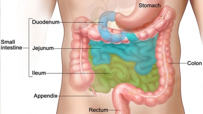
Duodenum
The first part of the small intestine is where bile and enzymes are added to the chyme to help break it down. It extends from the pyloric sphincter of the stomach, wraps around the head of the pancreas in a C-shape and ends at duodenojejunal flexure. The duodenum has four parts: superior (duodenal bulb/ampulla), descending, horizontal and ascending.
Jejunum
This is the middle section of the small intestine, where most of the digestion and absorption of food takes place. The jejunum is entirely intraperitoneal as the mesentery proper attaches it to the posterior abdominal wall.
Ileum
The final, longest section of the small intestine absorbs some nutrients. It is found in the lower right quadrant of the abdomen, although the terminal ileum can extend into the pelvic cavity. The ileum terminates at the ileal orifice (ileocecal junction) where the cecum of the large intestine begins.
lleocaecal valve
The ileocecal valve is a sphincter muscle situated at the junction of the ileum (last portion of your small intestine) and the colon (first portion of your large intestine). Its function is to allow digested food materials to pass from the small intestine into your large intestine. Chyme from the small intestine passes through here to the large intestine.
Looking inside
A cross-section through part of the small intestine shows the muscles that help to push food along its length. The lining is covered with millions of tiny finger-like projections called villi (a single projection is a villus).
Picture Credit : Google