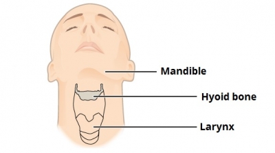
The hyoid bone is a ‘U’ shaped structure located in the anterior neck. It lies at the base of the mandible (approximately C3), where it acts as a site of attachment for the anterior neck muscles.
The hyoid consists of a body, two greater horns, and two lesser horns. The body forms the central quadrilateral-shaped broad segment of the hyoid. The greater horns are larger and longer than the lesser horns of the hyoid. The greater horns and lesser horns are also known as cornu majus and cornu minus, respectively. The body and the greater horns appear to give the hyoid its U-shape with the greater horns forming the limbs of the “U” on either side of the body. The greater and lesser horns normally unite to the body of the hyoid via fibrous tissue or a true joint. As age progresses, there is a physiological progression of ankylosis of the joints connecting the greater and lesser horns with the body of the hyoid.
The hyoid takes part in all possible functional actions of the orofacial complex. It preserves the patency of the airway between the oropharynx above and tracheal rings below. It also connects to the larynx and hence plays a role in phonation. Other functions include tongue movement, mastication, swallowing, prevention of regurgitation, and even respiration. Furthermore, the hyoid maintains the posture of the head, due to the complex connection it presumes between the mandible and the cervical spine.
Picture Credit : Google