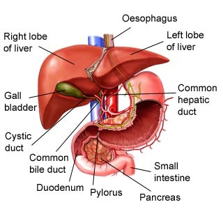
The liver is in the upper right abdomen, just below the diaphragm. It is divided into two lobes, or sections. An average adult liver is the size of a football and weighs 1.5 kg (3 lb).
Inferior vena cava
It is located at the posterior abdominal wall on the right side of the aorta. The IVC’s function is to carry the venous blood from the lower limbs and abdominopelvic region to the heart. This vein carries blood back to the heart.
Hepatic vein
Oxygen poor blood is drained from the liver to the inferior vena cava in this vein. The main hepatic veins are the right, intermediate and left hepatic veins. In addition, several smaller and somewhat inconsistent caudate lobe veins contribute to the venous drainage of the liver.
Hepatic duct
The common hepatic duct is the part of the biliary tract formed by the convergence of the right hepatic duct (which drains bile from the right functional lobe of the liver) and the left hepatic duct (which drains bile from the left functional lobe of the liver). This tube drains bile from the right lobe.
Tiny factories
The liver is made up of hexagonal (six-sided) units, called lobules. Each one is the size of a grain of sand. Blood flows from vessels in the lobule corners, is processed by the lobule’s cells, then is collected by the central vein and returned to the heart.
Left lobe
This is the smaller of the two liver lobes. When looking at the front of the liver, the left lobe of liver is divided from the right by the falciform ligament, which attaches the liver to the front wall of the body. The ligamentum venosum and ligamentum teres divide the left lobe of liver from the right as viewed from behind.
Right lobe
This is the biggest section of the liver. The right lobe liver has four sections. It is divided into the anterior right lobe and posterior right lobe by the right hepatic vein. It is also divided into the upper right lobe and lower right lobe by the portal vein.
Ligament
This connective tissue lies between the two main lobes. Liver ligaments are double-layered folds of peritoneum that attach the liver to surrounding organs, or to the abdominal wall. The majority of ligaments associated with the liver are remnants of embryological blood vessels that regressed as the fetus developed.
Hepatic artery
Oxygen-rich blood is supplied to the liver by this artery. It is the largest branch of the celiac trunk and the only one that courses to the right across the epigastric region of the abdomen. The common hepatic artery supplies blood to the liver, pylorus of the stomach, duodenum, pancreas, and gallbladder.
Cystic duct
This tube carries bile to and from the gallbladder. The cystic duct connects the top of the gallbladder’s neck to the common hepatic duct. It then joins the common bile duct, which meets pancreatic duct before it empties into the duodenum. In the average adult, the cystic duct measures four centimeters in length.
Gallbladder
The gallbladder stores bile made by the liver. The gallbladder is a small bag that stores bile it receives from the liver, concentrates it, then releases it into the duodenum. The gallbladder stores, processes, and releases bile, a liquid that helps the body to digest fat.
Worker lobules
Blood vessels feed the cells of the lobules, delivering oxygen-rich blood from the heart, and nutrients-rich blood from the small intestine. The cells inside the lobule extract and store the nutrients, release other vital chemicals into the blood, and make bile.
Picture Credit : Google