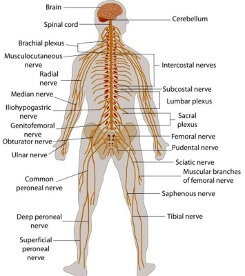
The brain and the spinal cord – the mass of nerves running down the backbone – make up the central nervous system (CNS). This coordinates most of the body’s activities, from blinking and breathing to seeing and standing. Nerves branch out to the rest of the body via the peripheral nervous system (PNS).
Brain
The brain is made of four parts; cerebrum, diencephalon, cerebellum and brainstem. Together these parts process the incoming information from peripheral tissues and generate commands; telling the tissues how to respond and function. These commands tackle the most complex voluntary and involuntary human body functions, from breathing to thinking. The control centre of the nervous system, this is home to more than 100 billion neurons.
Cranial nerves
Cranial nerves are peripheral nerves that emerge from the cranial nerve nuclei of the brainstem and spinal cord. They innervate the head and neck. Twelve pairs of cranial nerves relay signal between the brain and the head, face, and neck.
Spinal cord
The body’s primary communications highway, this carries all nerve signals between the body and brain. It also has the ability to generate commands but for involuntary processes only, i.e. reflexes. However, its main function is to pass information between the CNS and periphery.
Brachial plexus
The brachial plexus can be very challenging while studying anatomy. Even though it is essentially just a network or ‘bunch’ of nerves, it seems like it has very complex origin and branches, and students often get lost while reading the textbooks. This collection of nerves supplies the muscles and skin of the arm and hand.
Musculocutaneous nerve
The musculocutaneous nerve is responsible for very important function we use every day, bending/flexing our elbows. Tasks such as lifting a cup and brushing our teeth can become very difficult if the nerve is not functioning well. This nerve supplies muscles in the upper arm and gives feeling in the forearm.
Intercostal nerve
The intercostal nerves are the somatic nerves that arise from the anterior divisions of the thoracic spinal nerves from T1 to T11. These nerves in addition to supplying the thoracic wall also supply the pleura and peritoneum. The intercostals nerve supplies the muscles and skin of the thorax (chest).
Axillary nerve
One of the terminal branches of the brachial plexus is the axillary nerve, which is derived from the posterior cord (C5-6). It travels through the quadrangular space together with the posterior circumflex artery and vein. The axillary nerve supplies muscles and sensation in the shoulder.
Median nerve
The median nerve is the branch of the brachial plexus that supplies most of the superficial and deep flexors in the forearm, thenar and lumbrical muscles. It also gives sensation to certain areas of the skin of the hand. Most of the muscles in the forearm and hand, and some skin of the hand, is supplied by this nerve.
Phrenic nerve
The phrenic nerve is a bilateral, mixed nerve that originates from the cervical nerves in the neck and descends through the thorax to innervate the diaphragm. It is the only source of motor innervation to the diaphragm and therefore plays a crucial role in breathing. Messages to and from the diaphragm are carried by this nerve.
Radial nerve
The radial nerve is an essential component of the upper limb innervation network. It innervates essentially all the muscles on the posterior aspect of our arms, and is therefore a large nerve. This nerve supplies muscles in the back of the arm and the skin of the lower arm.
Lumbar plexus
A “plexus” is a branching network. The lumbar plexus supplies the skin and muscle of the lower back. The lower limbs have the tremendous responsibility of mobilizing and stabilizing the human body. Muscles of the pelvic region, posterior abdominal wall, and the fifty-nine muscles of the lower limb, as well as their corresponding joints, are innervated by branches of the lumbosacral plexus.
Ulnar nerve
The ulnar nerve can broadly be described as the nerve of the hand, as the nerve innervates the vast majority of the intrinsic hand muscles. It is one of the most clinically applicable nerves, due to its superficial course, and clinically apparent role in hand function. This article shall discuss the anatomy of the ulnar nerve, its precise course, as well as the clinical relevance it has. Supplying muscles in the forearm and hand, this nerve gives the ‘funny bone’ tingle if you knock your elbow.
Femoral nerve
The femoral nerve is a mixed nerve of the lower limb that innervates the muscles and skin of the hip and thigh. The femoral nerve originates from the lumbar plexus, from the ventral rami of L2-L4 spinal nerves. In fact, it is the longest branch of the lumbar plexus. The skin and muscle of the pelvis and leg are supplies by this web of nerves.
Sciatic nerve
The thickest and longest nerve in the body, this links the spinal cord to muscles in the legs and feet. The sciatic nerve starts as a collection of nerve fibers in the lower spine. These nerve fibers, or roots, exit the spinal canal through a number of openings in the bones at each level of the lower spine called foramina.
Saphenous nerve
The saphenous nerve is a sensory branch of the femoral nerve (lumbar plexus L3, L4), and supplies sensation to the anteromedial, medial and posteromedial surface of the leg. The saphenous nerve is the largest terminal cutaneous branch of the femoral nerve (dorsal divisions of the ventral rami of L2-L4).The saphenous nerve supplies the skin on the inner leg.
Common peroneal (fibular) nerve
The common fibular nerve, also known as the common peroneal nerve, is one of two main muscular branches of the sciatic nerve. A branch of the sciatic nerve, this supplies the front and side of the lower leg.
Superficial peroneal nerve
The superficial fibular nerve is a nerve of the lower limb. In older texts, it is known as the superficial peroneal nerve. Supplying the skin and muscles of the leg and foot, this is one of the fibular nerves.
Deep peroneal (fibular) nerve
The deep peroneal nerve, more commonly known as the deep fibular nerve innervates a number of muscles in the leg and foot, which are essential for normal gait and movement of the ankle. This nerve supplies the muscles of the leg and foot.
Tibial nerve
The tibial nerve is one of two main muscular branches of the sciatic nerve that innervates the triceps surae, plantaris, popliteus, tibialis posterior, flexor digitorum longus and flexor hallucis longus muscles. This is the biggest branch of the sciatic nerve. It produces a “pins” and “needles” feeling in the legs if it is squashed.
Plantar nerve
The plantar nerves are a pair of nerves innervating the sole of the foot. They arise from the posterior branch of the tibial nerve. The plantar nerve is responsible for the tickling sensation when the soles of the feet re touched.
Picture Credit : Google